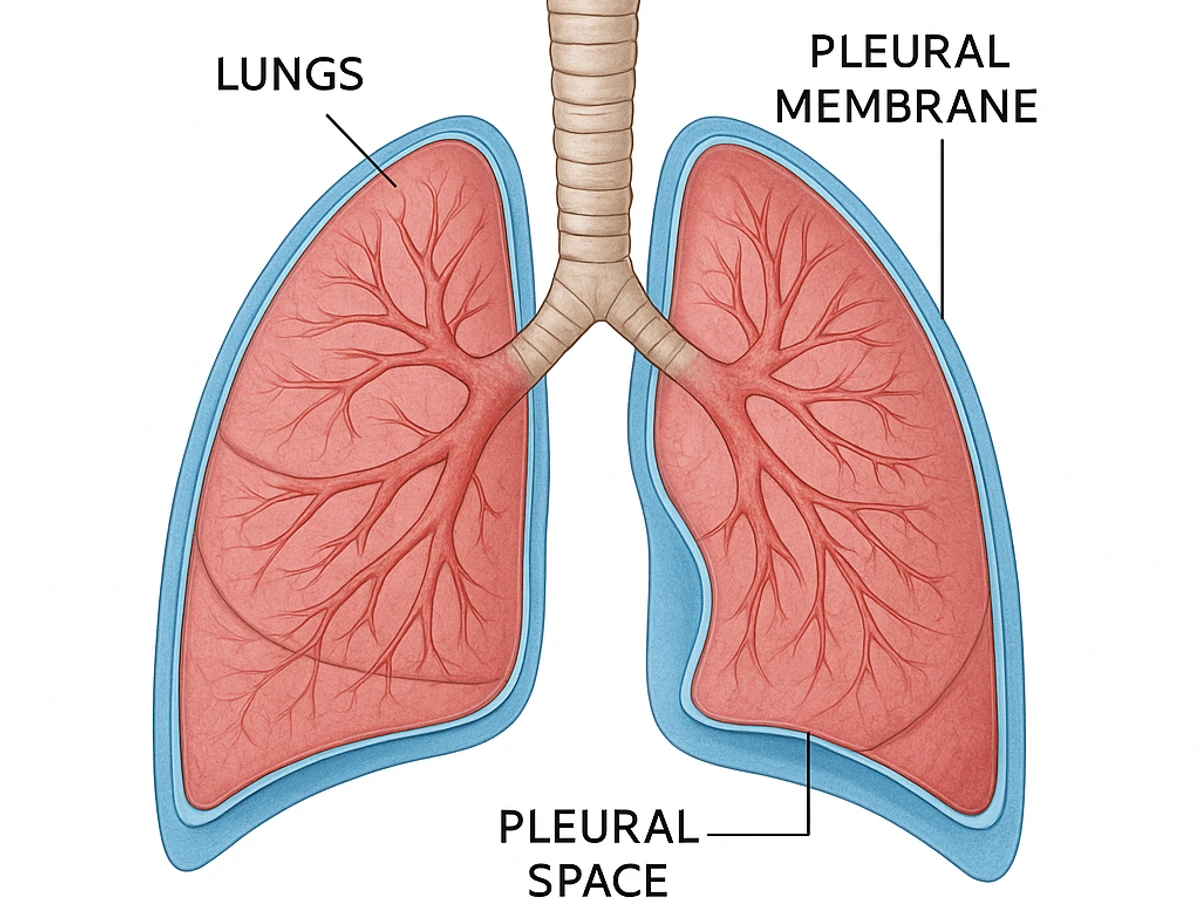
What is Pleural Effusion?
Pleural effusion is the abnormal accumulation of fluid in the pleural space—the area between the visceral pleura (the membrane lining the lungs) and the parietal pleura (the membrane lining the chest wall). In a healthy person, a small amount of clear fluid (about 10-20 mL) exists in this space to allow smooth movement of the lungs during breathing.
Fluid Composition in Mesothelioma
In pleural mesothelioma patients, the fluid becomes a pale yellow or blood-stained exudate containing inflammatory cells, cancer cells, and proteins. The increased production of this fluid is a hallmark of pleural mesothelioma and can range from minimal to several liters, significantly compromising lung function.
Why Mesothelioma Causes Fluid Buildup
The pleural effusion in mesothelioma results from several mechanisms working together:
Inflammatory Response
Mesothelioma tumors irritate the pleural membrane, triggering inflammation. This causes increased vascular permeability, allowing fluid and proteins to leak from blood vessels into the pleural space. The inflammation also stimulates cells to produce more fluid.
Lymphatic Obstruction
Cancer cells can block lymphatic vessels that normally drain fluid from the pleural space. This obstruction prevents normal fluid reabsorption, causing progressive accumulation over time.
Increased Vascular Permeability
Mesothelioma cells release substances that increase the permeability of blood vessel walls, allowing fluid to leak into the pleural cavity more readily than normal.
Symptoms of Pleural Effusion
The symptoms of pleural effusion in mesothelioma depend on the volume of fluid and how quickly it accumulates:
Common Symptoms
- Shortness of breath (dyspnea): The most common symptom, worsening with activity or when lying down
- Persistent cough: Dry or productive cough caused by lung compression
- Chest pain or pressure: Dull, aching pain that may worsen with deep breathing or coughing
- Fatigue: Reduced oxygen intake leads to tiredness and reduced exercise tolerance
- Decreased exercise tolerance: Inability to perform activities that were previously manageable
Severe or Rapid Onset
When large volumes of fluid accumulate rapidly, patients may experience acute onset of severe shortness of breath, lightheadedness, or chest discomfort requiring urgent medical attention.
Diagnosis and Imaging of Pleural Effusion
Pleural effusion is typically discovered during imaging performed for mesothelioma evaluation or symptom investigation.
Chest X-Ray
The primary imaging tool for detecting pleural effusion. Even small effusions (as little as 200 mL) can be visible on a standard chest X-ray. The fluid appears as a white opacity at the lung bases, often obscuring part of the diaphragm.
CT Scan
Provides more detailed information about effusion size, characteristics, and associated pleural thickening or tumors. CT helps determine the type of effusion and guide drainage procedures.
Ultrasound
Used to assess effusion characteristics and guide needle placement during thoracentesis. Ultrasound can determine if fluid is loculated (separated by membranes) and identify safe areas for needle insertion.
Thoracentesis for Analysis
When fluid is drained for symptom relief, the sample is analyzed for cell count, chemistry, protein levels, and presence of cancer cells. This analysis helps confirm mesothelioma diagnosis and provides prognostic information.
Drainage Procedures for Pleural Effusion
Several procedures can provide relief from pleural effusion symptoms:
Thoracentesis (Needle Aspiration)
Procedure: A needle is inserted through the chest wall, guided by imaging, and positioned in the pleural space. Fluid is withdrawn through the needle or catheter into sterile collection bottles.
- Duration: 10-20 minutes in an outpatient setting
- Recovery: Same-day discharge with brief observation
- Benefits: Quick symptomatic relief; fluid can be analyzed for diagnosis
- Limitations: Relief is often temporary; fluid typically reaccumulates within days to weeks
Chest Tube Placement
For patients with large effusions or when pleurodesis is planned, a chest tube (also called a thoracostomy tube) may be inserted through the chest wall. The tube remains in place, allowing continuous or intermittent drainage until pleurodesis takes effect or fluid production decreases.
PleurX Catheter (Tunneled Pleural Catheter)
A semi-permanent catheter designed for long-term home drainage management. The catheter is surgically placed in the pleural space with the external portion exiting the skin.
- Advantages: Allows home drainage without repeated hospital visits
- Frequency: Typically drained 2-3 times per week or as symptoms dictate
- Duration: Can remain in place for weeks to months
- Quality of life: Enables increased mobility and independence for patients
Pleurodesis: Preventing Fluid Reaccumulation
Pleurodesis is a procedure designed to permanently seal the pleural space and prevent fluid from reaccumulating. It involves introducing an irritant between the pleural layers to cause them to fuse together.
How Pleurodesis Works
The irritant substance triggers an inflammatory reaction in the pleura. This inflammation causes the visceral and parietal pleura to adhere together, essentially obliterating the pleural space where fluid would normally accumulate. Once successful, new fluid cannot collect in the sealed space.
Pleurodesis Agents
- Talc: Most effective agent with 80-90% success rates; typically delivered as a slurry or powder
- Doxycycline: Antibiotic that causes inflammation; success rate 60-70%
- Bleomycin: Chemotherapy agent; moderate success rate
- Iodopovidone: Antimicrobial agent with moderate effectiveness
Mechanical vs. Chemical Pleurodesis
Chemical pleurodesis uses irritant agents as described above. Mechanical pleurodesis involves physically abrading the pleural surfaces during surgery to encourage adhesion. Mechanical pleurodesis is more effective but requires surgical intervention.
Success Factors
Success depends on several factors: the agent used, lung re-expansion (the lung must expand fully to seal the space), absence of trapped lung (fluid preventing full expansion), and patient factors like inflammation status.
When to Seek Emergency Care
While pleural effusion is a chronic problem in mesothelioma, certain acute developments require immediate medical evaluation:
Critical Warning Signs
- Severe shortness of breath at rest that doesn't improve with usual measures
- Acute chest pain that radiates or is accompanied by dizziness
- Rapid or irregular heartbeat with breathing difficulty
- Confusion, severe dizziness, or loss of consciousness
- Coughing up blood or blood-tinged sputum
- High fever with chest pain or shortness of breath
- Inability to lie flat or speak in full sentences
Complications Requiring Urgent Care
These symptoms may indicate pneumonia, infection of the pleural space (empyema), collapsed lung, or heart involvement—all of which require immediate hospital-level evaluation and treatment.
Frequently Asked Questions About Pleural Effusion
What is pleural effusion in mesothelioma patients?
Pleural effusion is the buildup of fluid around the lungs between the two layers of the pleura. In mesothelioma patients, this occurs because cancer cells irritate the pleural membrane and cause inflammation, leading to excessive fluid production. This fluid accumulation can cause shortness of breath, chest pain, and reduced lung capacity.
What is thoracentesis and why is it performed?
Thoracentesis is a procedure where a needle is inserted through the chest wall to drain fluid from the pleural space around the lungs. It provides immediate relief from breathing difficulties and fluid pressure. The fluid removed can also be analyzed for cancer cells to help confirm a mesothelioma diagnosis. The relief is often temporary as fluid may reaccumulate.
How does pleurodesis work and is it permanent?
Pleurodesis is a procedure that seals the pleural space to prevent fluid reaccumulation. An irritant (such as talc or doxycycline) is introduced between the pleural layers, causing inflammation that bonds them together. This prevents fluid from accumulating in the space. Success rates vary, with about 60-90% of patients experiencing long-term relief, though some may require additional procedures.
What is a PleurX catheter and how is it used?
A PleurX catheter is a semi-permanent tunneled catheter placed in the pleural space that allows patients or caregivers to drain fluid at home using a collection bottle. This provides convenient, ongoing fluid management without repeated hospital visits. Patients typically drain fluid 2-3 times per week or as needed based on symptoms.
When should I seek emergency care for pleural effusion?
Seek emergency care if you experience severe shortness of breath at rest, chest pain that doesn't improve with drainage, rapid heartbeat, confusion or dizziness, coughing up blood, or signs of infection (fever, chills, drainage site redness). These may indicate complications requiring immediate medical attention.
Sources & References
Medically Reviewed
Dr. Sarah Chen, MD, MPH
Board-Certified Oncologist — Thoracic Oncology Specialist
Last reviewed: March 2026 | Our Editorial Process